
Key points
- Eczema is a chronic skin condition marked by chronic redness, dryness and itch, underpinned by a defective skin barrier function
- Management includes parent education and a daily skin care program.
- Early and liberal use of topical corticosteroids are recommended as has been shown to limit total corticosteroid dose.
- Superimposed infections are common and require prompt treatment.
- Emergency management of eczema often involves consideration of illness, managing infection, managing the acute impacts of itch (e.g. disturbed sleep) and making a decision on appropriate follow up arrangements.
- Moderate to severe eczema starting under 6 months of age should be considered for priority follow-up arrangements due to the ongoing skin disease with an associated higher likelihood of food allergy1
Purpose
This document provides clinical guidance for all staff involved in the care and management of a child presenting to an Emergency Department (ED) in Queensland with eczema.
This guideline has been developed by the Dermatology Service, Queensland Children’s Hospital, with input from senior ED clinicians and Paediatricians across Queensland. It has been endorsed for use across Queensland by the Queensland Emergency Care of Children Working Group in partnership with the Queensland Emergency Department Strategic Advisory Panel and the Healthcare Improvement Unit, Clinical Excellence Queensland.
Introduction
Eczema, also known as atopic dermatitis (AD), is a chronic inflammatory pruritic skin disease characterised by flare-ups and remissions.
Eczema is very common affecting 30% of Australian children.2,3 It is highly heritable with other family members often suffering from eczema or other atopic disorders (such as allergic rhinitis or asthma). In 60% of cases, the onset of eczema typically occurs in the first year of life (commonly at three to six months of age) with 90% developing symptoms by five years of age.2,4
Children with untreated moderate-to-severe eczema, under six months of age, go onto have a higher incidence of food allergies. For this reason, all causes of moderate-to-severe skin conditions that started under 6 months of age should be considered a priority when making follow up arrangements.
Eczema cannot be cured but can be effectively managed. The majority of children grow out of eczema by 16 years of age.
Pathogenesis
Eczema is thought to be caused by a complex interplay between genetic defects in skin barrier function, upregulation of inflammatory cytokines and environmental factors (such as second-hand smoke, climate, soaps).
Clinical presentation
While eczema normally affects the flexural surfaces (i.e. cubital fossa, popliteal fossa), the face, neck and extensor surfaces may be affected in children. The nose, groin and axillary regions are typically spared. Clinical presentation varies depending on the age, environment and ethnicity of the child.2,7,9
Superimposed bacterial or viral infections can occur due to the disordered barrier function and reduction in antimicrobial function of the skin. Bacterial infections are commonly caused by Staphylococcal aureus (impetiginized eczema) or Streptococcal species. Viral infections include eczema herpeticum (typically due to HSV type 1 or 2, presents 5-12 days after contact with an infected individual) and eczema coxsackium (enterovirus). Co-infections can occur.
| Age | Presentation |
|---|---|
Birth to 6 months | Lesions exudative erythematous weepy papules and plaques. Particularly on the face. Examples 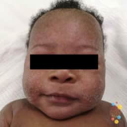 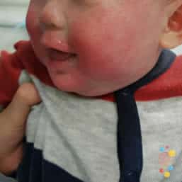 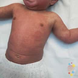 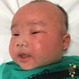  Images provided by Don’t Forget the Bubbles – Skin Deep https://dftbskindeep.com |
6 months to 12 years | Erythematous papules and plaques intermixed with lichenified plaques, often with erosions particularly in flexural areas. Examples 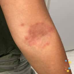  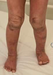 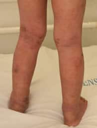 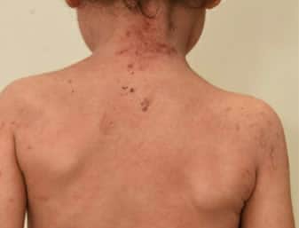 Images provided by Don’t Forget the Bubbles – Skin Deep https://dftbskindeep.com |
Over 12 years | Erythematous papules and plaques with xerotic scale and crust found on scalp, face, trunk, extensor surfaces or flexural surfaces. Lichenified plaques common in flexural or extensor surfaces depending on ethnicity. Examples 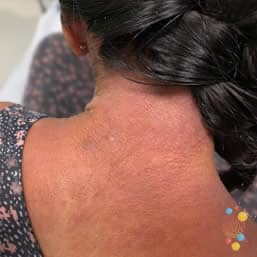 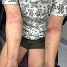 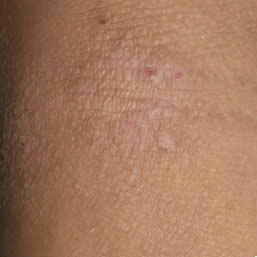 Images provided by Don’t Forget the Bubbles – Skin Deep https://dftbskindeep.com |
| Term | Description |
|---|---|
Papules |
Examples 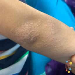 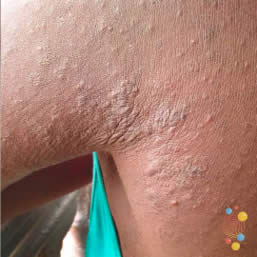 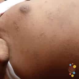 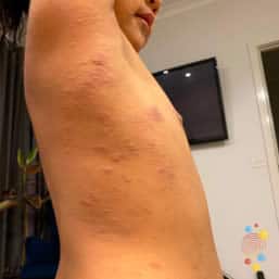 Images provided by Don’t Forget the Bubbles – Skin Deep https://dftbskindeep.com |
Lichenification | Palpably thickened skin with increased skin markings and lichenoid scale (caused by chronic rubbing). Example 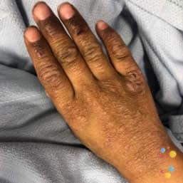 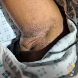  Images provided by Don’t Forget the Bubbles – Skin Deep https://dftbskindeep.com |
Discoid or nummular eczema |
Example 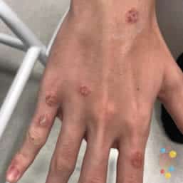  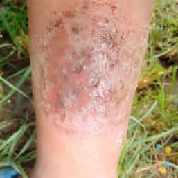 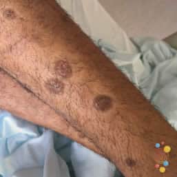 Images provided by Don’t Forget the Bubbles – Skin Deep https://dftbskindeep.com |
Post inflammatory hypo/hyperpigmentation (secondary to resolved eczema) | Hypo or hyper-pigmented macules in sites of resolved eczema. A response to inflammation; NOT due to topical cortisone use. Will resolve overtime, once eczema has not affected the area for at least 6 months. Example 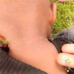 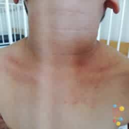 Images provided by Don’t Forget the Bubbles – Skin Deep https://dftbskindeep.com |
Assessment
The aim of the assessment is to:
- Identify children with a superimposed infection to enable appropriate treatment and, if required, specialist referral.
- Understand the extent and severity of disease to enable appropriate skincare management plan and need for specialist referral
- Identify moderate to severe eczema starting before 6 months of age and consider follow up targeted toward this group’s higher risk of food allergy.
History
History taking should include specific information on:
- Onset and duration of symptoms
- Effect on sleep (how much they wake due to itch) and number of days of school (or parents work) missed due to eczema
- Recent impact on quality of life and psychological well-being (e.g. restriction of activities, school absence, sleep disturbance)
- Recent symptoms suggestive of an infection. Please note severe skin disease is associated with temperature dysregulation and particularly a tendency toward hypothermia.
- Previous history/family history of atopy
- Usual topical skin care regimen
- Dietary and growth history – specifically ask about consumption and exposure to staple allergenic foods ( dairy, egg, wheat, soy , seafood, nuts, sesame)
Examination
A holistic approach to examination should be taken; consider both the severity of the child’s eczema and also, the impact it is having on psychosocial wellbeing.
| Severity | Skin and physical severity | Impact on quality of life and psychosocial wellbeing |
|---|---|---|
| Clear |
|
|
| Mild |
|
|
| Moderate |
|
|
| Severe |
|
|
Refer to the introduction for examples of papule, lichenification, discoid, nummular eczema and bullous impetigo.
Clinical features of superimposed infections
| Infection type | Description |
|---|---|
Impetiginised eczema |
Example 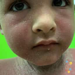 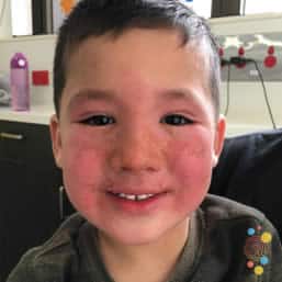 Images provided by Don’t Forget the Bubbles – Skin Deep https://dftbskindeep.com |
Eczema herpeticum |
Example 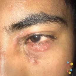 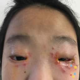 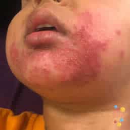 Images provided by Don’t Forget the Bubbles – Skin Deep https://dftbskindeep.com |
Eczema coxsackium |
Example 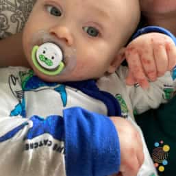 Images provided by Don’t Forget the Bubbles – Skin Deep https://dftbskindeep.com |
Bullous impetigo |
Example 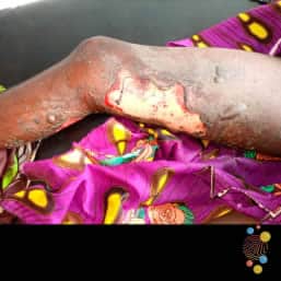  Images provided by Don’t Forget the Bubbles – Skin Deep https://dftbskindeep.com |
Investigations
Investigations such as lesion swabs are only indicated for children with suspected bacterial or viral infection. Request bacterial culture (m/c/s) and HSV PCR; VZV PCR if varicella is suspected.
If a viral skin infection is suspected in a child with blisters, rupture the blister and firmly swab the base of the lesion with a dry (no medium) swab.
Seek senior paediatric or dermatology advice for children with moderate to severe disease. Ophthalmology advice either locally or via CATCH should be sought for suspected viral infections involving or possibly involving the ophthalmic branch of the trigeminal nerve.
Differential diagnoses for eczematous lesions
- Infantile seborrhoeic dermatitis (generally not itchy)
- Tinea (dermatophyte)
- Scabies (sudden onset of burrows, papules and pustules to palms, soles and webbing in non-atopic child with family history of pruritus, papular pruritic rash to torso and limbs may follow)
- Periorificial dermatitis (small erythematous papules around mouth, nose, and eyes, exacerbated by moderate or potent steroids)
- Psoriasis (itchy, well-defined, salmon pink, silvery scaly plaques on extensor surfaces of elbows, knees, and also scalp)
- Miliaria
- Zinc deficiency/other nutritional deficiency
- Other
Management
Seek senior emergency/paediatric advice for a child with mild to moderate eczema who is febrile or unwell. The mainstay of eczema management revolves around education and development of a skincare management plan.
Education
Parents/carers should receive education regarding:
- Eczema symptoms (xerosis and inflammation), come and go in a cyclical fashion.
- Brief pathophysiology of disordered barrier function leading to increase permeability (water loss), reduction of natural moisturising factors, decreased microbial function, and immune dysregulation (over-active immune response to the environment).
- Need for an everyday skincare management plan (to minimise xerosis and therefore pruritus) and a flare management plan to control flares quickly, improving sleep and quality of life.
- Role of allergens (such as house dust mite, animal dander, and pollens) – may be contributory but do not cause eczema.
- Food allergies coexist in 10-20% of children with eczema10-12 however, they are not the cause of eczema. It is important that the child is still exposed to all allergenic foods. Any concern of immediate allergic symptoms (e.g. urticaria or angioedema, GI symptoms or signs of anaphylaxis) may be referred to General Paediatrics or Immunologist/Allergist.
- Flares associated with food
- require clear and reliable signs of a significant flare within two hours of food ingestion
- different to a food allergy
- providing the flares can be managed, the food can remain in the diet
- perioral rash in babies and toddlers after feeding could be an irritant eczematous rash from the contact of food to the skin and may not be an allergy
- Please see link https://preventallergies.org.au/eczema/eczema-and-food-allergy/ for further information.
- Topical corticosteroids
- Early, liberal use is recommended as this limits the total dose given over time.
- Use daily until the eczema is clear.
- Side effects (thinning of the skin and telangiectasia) are usually only associated with inappropriate use e.g. daily use of potent corticosteroids, on normal skin, over many months.
- Hypopigmentation or hyperpigmentation is NOT a side effect of topical corticosteroids, rather a temporary result of the inflammatory process of eczema. This is not scarring. Pigment will normalise over time.
Skincare management plan
View: Eczema Action Plan [DOCM 98.27 KB]
Everyday skincare treatment
- Daily bath (or shower for teenagers) with a soap substitute (e.g. aqueous cream cleanser or soap free wash/bath oil)
- For very young infants, bathing just once or twice per week is appropriate
- Consider twice weekly antibacterial baths Refer to Eczema: Baths and video for further information.
Potassium permanganate (Condy’s Crystals) if prone to regular infections.
Add a very small amount to bath to make water PALE PINK in colour.The colour is best determined in a white bath. If this is not possible, scoop out some solution in a clear jug to determine colour.
Soak child for approx. 10 minutes. Use a face washer to wash face with solution **Caution: This over-the-counter product is corrosive when undiluted. Avoid contact of the crystals or strong solutions with the eyes, mouth, nose and other mucous membranes.
- Bleach baths8 and salt if prone to regular flares
Add 12mL of sodium hypochlorite 4% bleach and 1/3 of a cup of pool or table salt to 10L water. Measure how many 10L buckets fill the bath to the desired level and write this down. Add 12mL of sodium hypochlorite 4% bleach for every 10L water. Add 1/3 cup of pool or table salt for every 10L water. **Caution: These over-the-counter products can be corrosive when undiluted. Avoid contact with the eyes, mouth, nose and other mucous membranes. Use caution to avoid bleaching of clothing and surfaces with undiluted bleach.
If access to a bath is limited OR only a small area of the body is affected, localised soaks to the area can be done. A bucket filled with either potassium permanganate or bleach an salt solution listed above can be used; soak a cloth in the solution and hold onto the affected areas. Repeat for 5-10 minutes.
- Regular moisturiser head-to-toe (from two to six times daily)9,10
- Ointments are preferred over creams where tolerated.
- Creams will often sting very dry skin or broken skin.
- Avoid “natural”, animal, plant-based moisturisers or moisturises with food products e.g., nuts
- Avoid irritants/triggers: soap, frequent shampooing of hair and, where possible, heat, and sweat.
- For infants/toddlers: Minimise skin contact with saliva and foods.
- Wipe face regularly with wet cloth to remove saliva/food and apply thick barrier emollient.
- Mineral based sunscreens are preferred as they are less likely to irritate the child’s skin. These include Zinc or titanium dioxide as ingredients. Chemical blockers are more likely to irritate the skin with eczema.
- For infants/toddlers: Minimise skin contact with saliva and foods.
Flare treatment
- Continue everyday skincare treatment as per above
- Add daily antibacterial or anti-inflammatory baths daily for moderate to severe flares – Refer to Eczema: Baths and video for further information. Continue for 2 weeks then reduce to maintenance twice weekly as above.
- Topical corticosteroids (in line with disease severity) are recommended for once daily administration to eczematous areas until clear. The order of application of moisturiser and corticosteroid does not affect efficacy.
- There is little evidence twice or thrice daily application is more beneficial; for those who are admitted with severe exacerbation of eczema, twice daily application of topical corticosteroid for the duration of admission is often prescribed. Inpatient care is focused on skin condition optimization.
- There is a theoretical risk that topical corticosteroid applied over HSV vesicles may result in worsening infection. However, in most cases, controlling the underlying eczema with topical corticosteroids is more effective in preventing the spread of HSV.
- Topical corticosteroids should be used on raw and excoriated areas, even where bacterial superficial infection is suspected or proven.
Topical corticosteroids – Ointments vs creams vs lotions
The efficacy of the medication changes depending on the vehicle it is in. The most effective regime is one that is tolerated by the patient and family. This will be different for each child, and is often a combination different vehicles (e.g. ointment in worst areas/broken skin/at night when not hot and cream/lotion in other areas/ for maintenance/ during hotter times of the day).
- Ointments (oil/lipid base with no water) are preferred as they are more occlusive and are more effective at delivering the medication into the skin. They also provide an effective skin barrier to reduce water loss. Skin water loss impairs delivery of treatments into the skin.
- Creams (oil and water base, emulsified) and lotions (thinner emulsified oil and water base or alcohol based) may be appropriate for the scalp or very hairy areas.
- Creams may sting when applied to broken skin
- Lotions can contain alcohol and often sting broken skin, so they may not be well tolerated. Creams are recommended if lotion is not able to be tolerated in the child with an excoriated scalp for example
- Ointments reduce trans-epidermal water loss which also impairs heat loss. Ointments can cause overheating.
- For moderate to severe eczema add wet wraps/garments. Refer to Eczema: Wet Wraps.
| A fingertip unit (FTU) is from distal phalanx to tip of finger of an adult hand. A single FTU of topical corticosteroid will cover the equivalent of two adult hands of eczema and is equivalent to 0.5 g of topical corticosteroid. Calculate the number of grams required per application to determine how many tubes are required per script. There are PBS streamline authority codes for prescription of multiple tubes. |
| Potency | Active Ingredient | Use |
|---|---|---|
| Class I (mild) |
LAM Listed: Hydrocortisone acetate – cream or ointment 1% (eg Sigmacort / Cortic-DS brands) 30g sizeNon LAM listed options for supply from outside QH:
| Mild face / neck / genital inflammation |
| Class II (moderate) |
LAM Listed: Triamcinolone 0.02%
Betamethasone valerate 0.03% and clioquinol 0.9% (anti-microbial) in Aqueous Cream, 50g, QH Compounded
|
Mild to moderate body and scalp inflammation
Moderate/severe face / neck / genital inflammation Mild to moderate body and scalp eczema with excoriations; minimize the risk of superficial infection |
| Class III (potent) |
LAM listed: Betamethasone dipropionate 0.05% (Plain base)
Mometasone Furoate 0.1%
| Moderate to severe body and scalp inflammation |
| Class IV (very potent) |
LAM Listed:
Non LAM Listed (IPA required or organise for supply from outside QH:
Clobetasol propionate 0.05%
|
Severe inflammation Dermatologist only |
Treatment of superimposed infections
Bacterial infection
Oral antibiotics are recommended following a lesion swab.
Oral antibiotic dosing for the treatment of superimposed bacterial infection in child with eczema (dosing for infants and children > 1 month of age with normal renal function and appropriate allergy history).
Refer to CHQ Antibiocard for more information – Children’s Health Queensland Paediatric Antibiocard: Empirical Antibiotic Guidelines [PDF 710.4 KB]
Viral infection
Antiviral dosing for the treatment of superimposed herpetic infection in child with eczema (dosing for infants and children > 1 month of age with normal renal function).
| Valaciclovir (oral) |
20 mg/kg every eight hours (up to a 1g) for 7 days Oral preferred route. Good oral bioavailability. ID approval required |
| Aciclovir (IV) |
If systemically unwell and unable to tolerate oral therapy: Aciclovir IV 10 mg/kg (up to 500 mg) every eight hours for seven days only if able to be commenced within 72 hours of the appearance of the first lesion. ID approval required |
Treatments NOT recommended
- Avoid topical antibiotics (i.e. Bactroban, mupirocin) as this is less effective and may contribute to antibiotic resistance in moderate to severe cases.
- Antihistamines – may be useful for allergic rhinitis but do not significantly decrease the itch associated with eczema (eczema is not histamine related).
- Restrictive diets.
Escalation and advice outside of ED
Clinicians can contact the services below if escalation of care outside of senior clinicians within the ED is needed, as per local practices. Transfer is recommended if the child requires a higher level of care.
| Reason for contact | Who to contact |
|---|---|
Advice | Follow local practice. Options:
|
| Referral | The first point of call is usually the onsite/local paediatric service. Paediatric Dermatology services, e.g. Queensland Children’s Hospital, will accept referral’s for the following indications:
Consider referral to paediatric allergy/immunology service if food allergy is suspected with signs or concern of allergic reaction. Consider eczema education independent of specialist referral. QCH Dermatology Service offer 40 minute live web based group eczema education sessions for families and clinicians to the state of QLD (run 4 weekly on a Wednesday). If the child does not require referral to QCH Dermatology Service, but just education, please email DermatologyCN_QCH@health.qld.gov.au with patient details. The QCH Dermatology administration team will contact the family with the virtual appointment. |
Inter-hospital transfers
| Do I need a critical transfer? |
|
| Request a non-critical inter-hospital transfer |
|
| Non-critical transfer forms |
|
Disposition
When to consider discharge
On discharge, parent/carers should be provided with the following:
- Eczema action plan [DOCM 98.27 KB]
- Education on eczema and how to apply wet wraps (if required)
- Including watching video demonstration of wet wraps/eczema baths prior to discharge
- Scripts/supply of medications for treatment of eczema
Follow-up
- GP follow-up is required for all cases within 1-2 weeks
- Paediatric or Dermatology follow-up for moderate to severe eczema
- Specialist paediatric Allergy/Immunology if allergic reactions are suspected
- Virtual eczema education: once off 40-minute sessions. This program is run by the QCH dermatology nursing and administration teams
When to consider admission
Consider admission for:
- Moderate to severe eczema not responding to appropriate treatment
- Widespread erythematous / inflamed skin associated with thermoregulation dysfunction
- Disseminated eczema herpeticum
- Severely infected eczema
- Parental distress
- Moderate to severe impact on quality of life (e.g. significant sleep disturbance or school absence)
Related documents
Forms and factsheets
Videos
- Martin PE, Eckert JK, Koplin JJ, Lowe AJ, Gurrin LC, Dharmage SC, et al. Which infants are at risk of food allergy? Results from a population-based cohort. Clinical & Experimental Allergy, 2014; 45, 255-264.
- Eichenfield LF, Tom WL, Chamlin SL, Feldman SR, Hanifin JM, Simpson EL, et al. Guidelines of care for the management of atopic dermatitis: section 1. Diagnosis and assessment of atopic dermatitis. J Am Acad Dermatol. 2014;70(2):338-51.
- Mooney E, Rademaker M, Dailey R, Daniel BS, Drummond C, Fischer G, et al. Adverse effects of topical corticosteroids in paediatric eczema: Australasian consensus statement. Australas J Dermatol. 2015;56(4):241-51.
- Eichenfield LF, Totri C. Optimizing outcomes for paediatric atopic dermatitis. Br J Dermatol. 2014;170 Suppl 1:31-7.
- Beal BT, Prodanovic E, Kuo JE, Armbrecht ES, Peter JR, Siegfried EC. Impact of a Pediatric Dermatology Service on Emergency Department Utilization for Children with Dermatitis. Pediatr Dermatol. 2016;33(1):69-74.
- Pride HB, Tollefson M, Silverman R. What’s new in pediatric dermatology?: part II. Treatment. J Am Acad Dermatol. 2013;68(6):899 e1-11; quiz 910-2.
- Huang JT, Abrams M, Tlougan B, Rademaker A, Paller AS. Treatment of Staphylocoocus aureus colonization in atopic dermatitis decreases disease severity. Pediatrics. 2009; 123(5):e808-14.
- Pride HB, Tollefson M, Silverman R. What’s new in pediatric dermatology?: part I. Diagnosis and pathogenesis. J Am Acad Dermatol. 2013;68(6):885 e1-12; quiz 97-8.
- Hon KL, Leung AK, Barankin B. Barrier repair therapy in atopic dermatitis: an overview. Am J Clin Dermatol. 2013;14(5):389-99.
- Horimukai K, Morita K, Narita M et al. Application of moisturizer to neonates prevents development of atopic dermatitis. Journal of Allergy and Clinical Immunology. 2014;134:824-830
- Werfel T, Breuer K Role of food allergy in atopic dermatitis. Current Opinion in Allergy and Clinical Immunology. 2004;4(5):379-385
- Worth A, Sheikh A Food allergy and atopic eczema. Current Opinion in Allergy and Clinical Immunology. 2010; 10(3):226-230
- Martin PE, Eckert JK, Koplin JJ et al. Which infants with eczema are at risk of food allergy? Results from a population-based cohort. Clinical & Experimental Allergy. 2015; 45(1):255-264
Document ID: CHQ-GDL-60034
Version number: 1.0
Supersedes: New
Approval date: 27/04/2022
Effective date: 27/04/2022
Review date: 27/04/2026
Executive sponsor: Executive Director Medical Services
Author/custodian: Queensland Emergency Care Children Working Group
Applicable to: Queensland Health medical and nursing staff
Document source: Internal (QHEPS) + External
Authorisation: Executive Director Clinical Services
Keywords: Eczema, dermatology, steroids, herpeticum, bullous, impetigo, atopy, 60034
Accreditation references: NSQHS Standards: 1, 3, 4
This guideline is intended as a guide and provided for information purposes only. View full disclaimer.